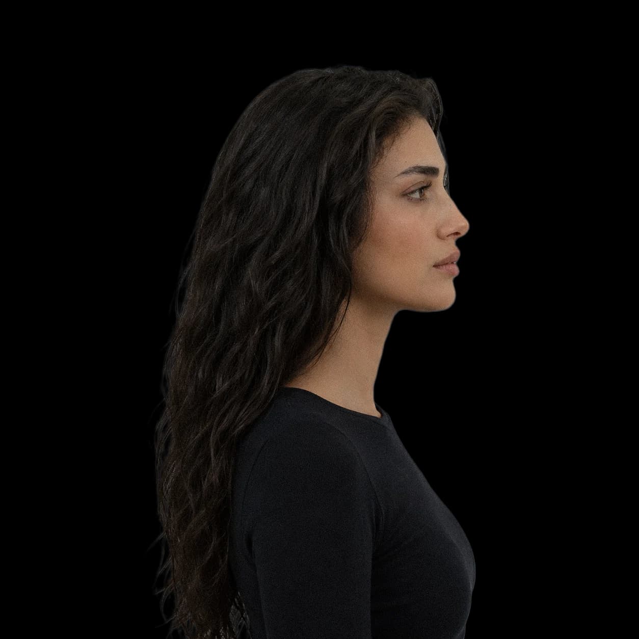
A recessed maxilla is an upper jaw that sits further back than normal relative to the skull base. It shows in profile as a flat area beside the nose, a nose base and upper lip with little support, and cheek bones that look set back. A forward maxilla projects the midface: the upper lip is supported, the area beside the nose is full, and the face has depth from the front. This article covers how forward position is measured, what pushes the maxilla back during growth, and how the sutures around it respond to mechanical force, with the geometry and the loading data worked through.
Landmarks
- SNA angle
- The angle between the sella-nasion line of the skull base and point A, the deepest point at the front of the upper jaw. Lower means a more recessed maxilla.
- Point A
- The deepest point on the front curve of the maxilla between the nose base and the upper incisor roots.
- Circummaxillary sutures
- The fibrous joints that hold the maxilla to the surrounding bones: frontomaxillary, nasomaxillary, zygomaticomaxillary, zygomaticotemporal and pterygopalatine.
- Microstrainµε
- Deformation of one millionth of a length. 1,500 µε means the tissue is compressed or stretched by 0.15%.
- Class III
- A bite in which the lower teeth sit forward of the upper teeth. A recessed maxilla is one of its causes.
What a recessed maxilla is
The maxilla forms the upper jaw, the palate, the floor of the nose and the base of the cheeks. Its forward position sets the support for everything in front of it: the upper lip, the base of the nose, and the soft tissue beside the nose. When it sits back, those tissues have less to rest on, and the middle of the face reads flat in profile. When it sits forward, the midface has depth and the lower face looks balanced beneath it.
A recessed maxilla is one cause of a Class III bite, where the lower teeth sit in front of the upper. Worldwide, Class III occurs in about 5.93% of the permanent dentition and 4% of the mixed dentition, with a range from 1% to 20% between populations and the highest rates in East Asian children (PMID 30672991). Not every recessed maxilla produces a Class III bite, because the lower jaw can sit back as well.
How forward position is measured, and what a degree is worth
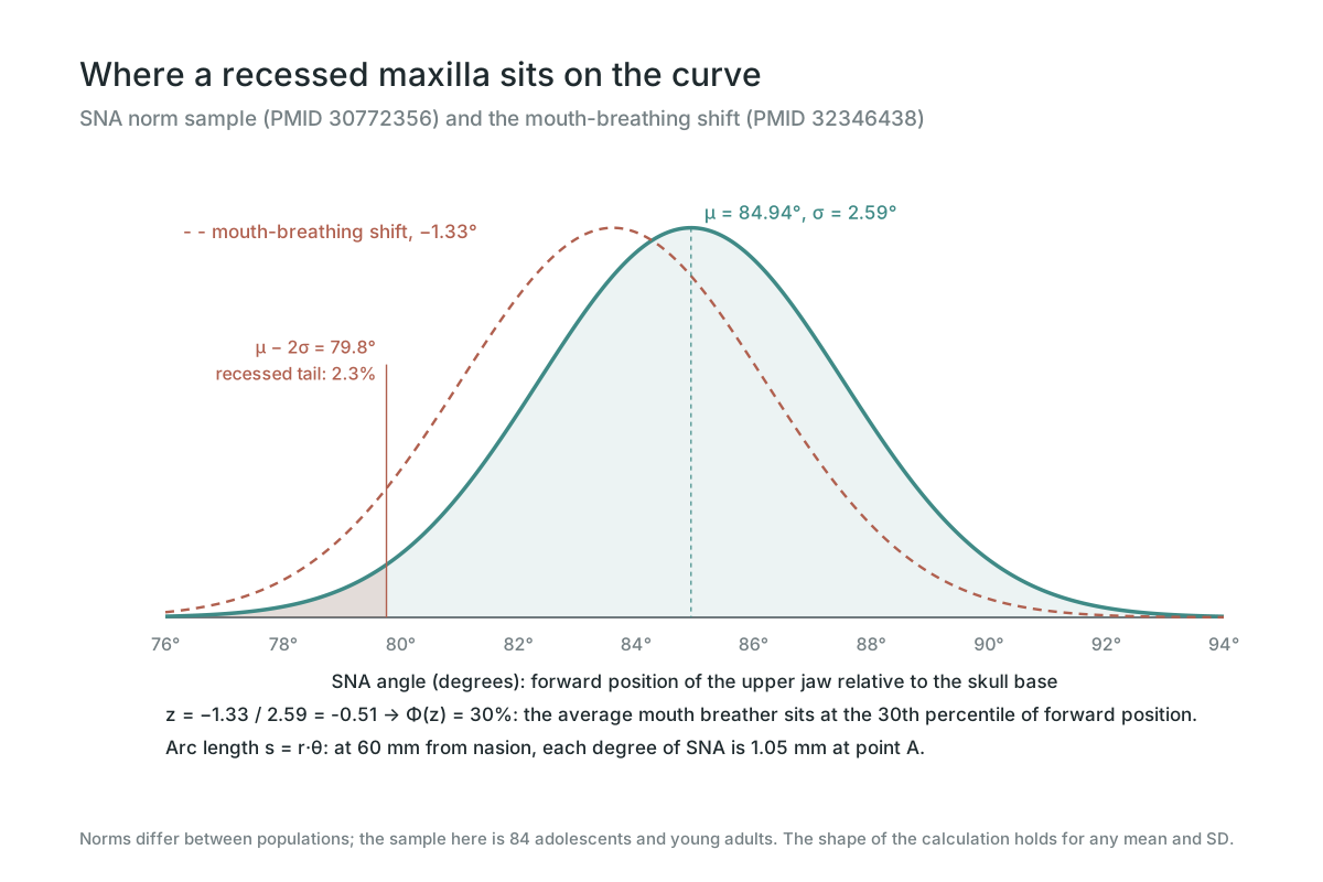
Cephalometrics measures maxillary position with SNA, the angle at nasion between the skull base line and point A. Norms depend on the population. In one published sample of 84 adolescents and young adults, SNA averaged 84.94° ± 2.59° (PMID 30772356). Treating SNA as normally distributed, the recessed tail below μ − 2σ = 79.8° holds Φ(−2) = 2.3% of people.
Degrees convert to millimetres by arc length, s = r·θ with θ in radians. Point A lies roughly 60 mm from nasion, so each degree of SNA is 60 × π/180 = 1.05 mm at the front of the upper jaw. A maxilla 3° behind the mean sits about 3 mm further back at its base, and the upper lip and nose base lose that much support.
The mouth-breathing difference fits on the same curve. With a shift of −1.33° against an SD of 2.59°, z = −0.51 and Φ(z) = 0.30: the average mouth breather sits at the 30th percentile of forward position rather than the 50th.
5.93%
Class III prevalence, permanent dentition
1.05 mm
Point A shift per degree of SNA
30th
Percentile of the average mouth breather
What sets the maxilla back
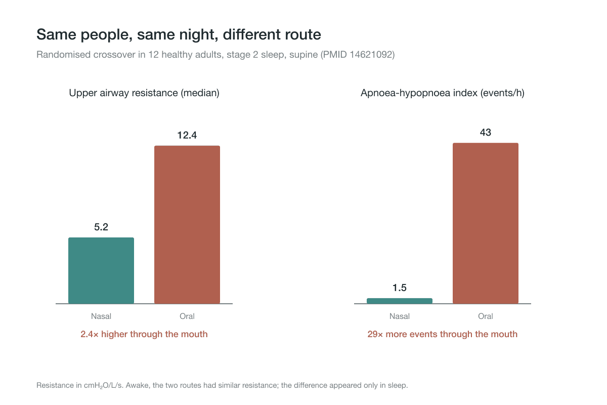
The strongest measured driver is breathing. A meta-analysis of 18 cephalometric studies found SNA 1.33° lower (95% CI −2.03 to −0.63) and SNB 1.33° lower in children who breathe through the mouth, with a longer lower face and a steeper mandibular plane (PMID 32346438). In primates, blocking the nose alone changed the face and bite of every experimental animal (PMID 6939331). The details are in mouth breathing face.
The same pattern shows up in adults with sleep apnoea. A meta-analysis of cephalometric studies found a maxillary length (ANS-PNS) 1.76 mm shorter, SNB 1.49° lower, a longer lower face and a narrower pharyngeal airway than in controls (PMID 27039222). Chewing load and arch width feed into it too; a narrow, underloaded maxilla is covered in narrow maxilla vs wide maxilla.
How the maxilla grows forward: sutures and their stem cells
The maxilla is not fused to the skull. It is held by a ring of sutures, fibrous joints with bone-forming cells along their edges. During growth, the maxilla is carried forward and down while bone is laid down at these sutures behind and above it. A suture is a growth site, and its cells read mechanical force.
Those cells include resident stem cells. In the zygomaticomaxillary suture, the joint between the maxilla and the cheek bone, Gli1⁺ cells proliferated under distraction force and co-localised with the bone transcription factor Runx2. Their primary cilia sensed the force and switched on Hedgehog signalling, which was required for the move toward bone (PMID 38971766). Across the midpalatal suture, tension shifted a subset of suture stem cells from a cartilage trajectory onto a bone trajectory, through a translational pathway involving Dalrd3 and Id3 (PMID 40528244).
The force sensor on the cell membrane is PIEZO1, a channel that opens when the membrane stretches and lets calcium in (PMID 34208464). Under tension, periosteal stem cells raised Runx2, Osterix and alkaline phosphatase through Piezo1 and CaMKII, and blocking Piezo1 reduced those signals (PMID 39350680). The biochemistry is a chain: stretch opens PIEZO1, Ca²⁺ enters, CaMKII is activated, and the osteogenic transcription factors that direct bone formation are turned on.
Age does not remove the cells. Human calvarial sutures contain mesenchymal stromal cells marked by GLI1 and AXIN2 (PMID 32575385). In adult mice, mechanical expansion of a functionally closed suture significantly increased its skeletal stem cells, and a critical-size bone defect created at the same time regenerated fully through Wnt signalling (PMID 37040407). Controlled force reopened a growth process that had gone quiet.
Static vs cyclic force at the suture
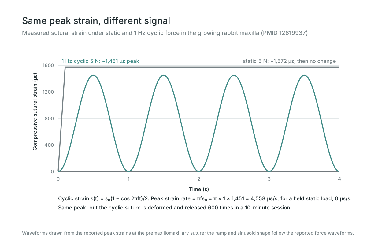
Kopher and Mao applied 5 N to the maxilla of growing rabbits, either as a static hold or as a 1 Hz cycle, and recorded the strain inside the sutures. The peaks were almost the same: −1,572 µε static and −1,451 µε cyclic at the premaxillomaxillary suture, which was compressed, and 134 vs 124 µε at the nasofrontal suture, which was stretched (PMID 12619937). Different force waveforms produce matching strain waveforms in the suture (PMID 14582615).
The mathematical difference is the rate. A cyclic strain ε(t) = ε₀(1 − cos 2πft)/2 has a peak rate of πfε₀ = π × 1 × 1,451 ≈ 4,558 µε/s, and it repeats: 600 load-release cycles in a 10-minute session. A held static load has a strain rate of zero once it is applied. Cells respond to change, and cyclic loading supplies change 600 times a session.
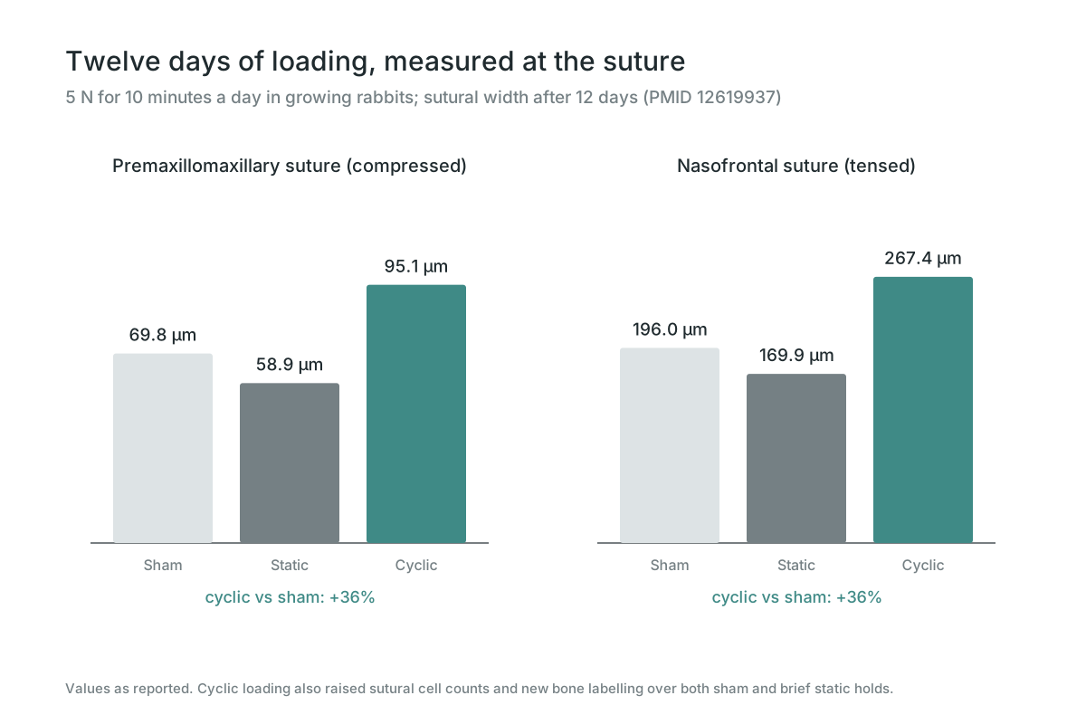
After 10 minutes a day for 12 days, cyclic loading widened the premaxillomaxillary suture to 95.1 µm against 69.8 in sham controls, and the nasofrontal suture to 267.4 µm against 196.0, both 36% more. It also raised sutural cell counts and new bone labelling. Brief static holds at the same peak (58.9 and 169.9 µm) did not exceed sham (PMID 12619937). Cyclic 1 N loading at 8 Hz, 20 minutes a day, likewise increased sutural width, cell density and osteoblast-lined bone surface, in both tension and compression (PMID 18032124). Mao's review puts the effective dose as low as 600 cycles a day (PMID 12454093).
Static force works when it is sustained. Every stem cell result above came from continuous expansion or distraction held for days, not from minutes-long holds. The two patterns do different jobs: sustained tension keeps a suture open and recruits its stem cells, and cyclic loading multiplies the growth signal on top.
Suture traction and thumbpulling
Manual suture work applies the same principle by hand. A Delphi expert process developed a cranial suture traction therapy sequence for facial asymmetry (PMID 35888588). Thumbpulling loads the maxilla from the palate, outward and slightly forward, and can be done as a static hold or in cycles. The anatomy, loading patterns and safety stops are in what is thumbpulling. The loading experiments in this article were done in growing animals; the millimetres an adult gains from manual traction have not been measured in a trial.
Why we treat forward growth as trainable
The maxilla is a bone held by living sutures, and those sutures still carry stem cells that answer to tension. That is why we treat forward growth as trainable. Breathe through the nose, load the palate and the midface sutures with steady traction, and add cyclic loading on top, because the controlled data show that the oscillating signal is what multiplies sutural growth.
Recessed vs forward maxilla, side by side
Each row is a measured value or a direct calculation from one.
| Measure | Recessed maxilla | Forward maxilla |
|---|---|---|
| SNA (norm sample) | Below 79.8° (μ − 2σ) | Around 84.9° or above |
| Point A per degree | About 1 mm further back | About 1 mm further forward |
| Midface profile | Flat beside the nose, less lip support | Full, supported upper lip and nose base |
| Typical bite | Tendency to Class III | Upper teeth in front of lower |
| Breathing link | SNA 1.33° lower in mouth breathers | Nasal breathing reference |
| Maxillary length in sleep apnoea | 1.76 mm shorter | Control reference |
Frequently Asked Questions
An upper jaw positioned further back than normal relative to the skull base, measured by a low SNA angle. It shows as a flat area beside the nose and less support for the upper lip and nose base.