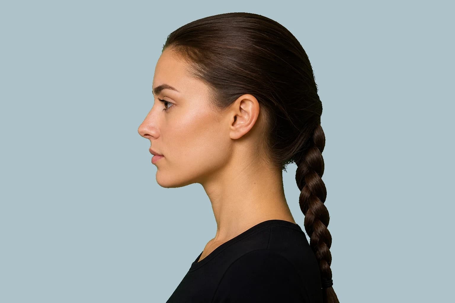
Nasion is a bony landmark at the junction of the frontal and nasal bones. Radix is the visible nasal-root region around it. A photograph shows the surface contour, including skin and soft tissue, rather than the suture itself. Human studies document nasal changes during growth and aging. Animal and cell experiments show responses to controlled mechanical loading, but they do not establish that hand pressure increases adult radix height.

Landmarks
- NasionN
- The bony point in the middle of the frontonasal suture, where the frontal bone meets the nasal bones. On a side X-ray, it is the deepest bony point at the nasal root.
- Radix
- The visible root of the nose. Its apparent height and depth depend on the nasal bones, forehead shape, skin thickness, fat, lighting, and camera angle.
- Frontonasal suture
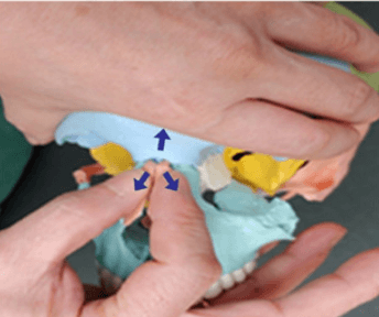
- The joint between the frontal bone and the nasal bones. Traction at nasion is intended to load this joint. A photograph shows the surface over it, not the suture itself.
- Nasofrontal angle
- The angle in a side view formed by points on the forehead or glabella, nasion, and the nasal bridge or tip. The measured value depends on which landmarks are used.
- Suture Traction TherapySTT
- Manual traction across facial sutures before palatal work. At the nasal root, the technique supports the frontal bone while applying force at nasion against the periosteum.
- Mechanotransduction
- The process by which cells turn physical force into biological signals. In sutures, this involves primary cilia, ion channels such as Piezo1, growth-factor pathways, and transcription factors such as Runx2 that regulate bone formation.
- Osteogenic differentiation
- The process by which a cell develops into a bone-forming cell. Signs include Runx2 and Osterix activity, alkaline phosphatase, osteocalcin, and mineral deposition.
Where are nasion and radix?
The bony nasion lies at the midline frontonasal junction. Soft tissue nasion is the deepest visible point of the nasal root in a profile image. The two should be labelled separately because the surface can obscure the exact bony location.
Radix describes the root region rather than one mathematical point. Its visible height and depth depend on the forehead, nasal bones and covering soft tissue. A low-looking root can reflect its vertical position, its forward projection or the surrounding contour.
The nasofrontal angle joins a forehead reference with a nasal reference at the root. State those lines: an angle measured toward the tip need not equal an angle using a short upper-bridge tangent.
- Bony landmark: nasion, at the frontonasal suture.
- Visible shape: radix, around the start of the nasal bridge.
- Angle: the nasofrontal angle, measured using the forehead, nasion, and nose.
- Photographs: shadows can change the apparent shape without showing what is happening at the suture.
What human growth and aging studies show
A longitudinal CBCT-derived cephalogram study of 30 untreated children found that nasion moved forward and downward relative to the cranial base. A separate series followed 40 children from ages 6 to 18 and documented changes in nasal and other soft tissue dimensions. These are growth observations in children (PMID 34960576, PMID 23834271).
Cross-sectional studies describe differences between people of different ages: one measured external nasal landmarks in 859 people aged 4–73, while another used CT data from 321 people spanning infancy to adulthood. They help describe developmental patterns but do not track an intervention in one person (PMID 20729017, PMID 34549897).
Paired CT images of 48 Korean adults approximately 12 years apart showed increased nasal length, bridge height and nasofrontal angle on average. Individual variation was substantial. Aging changes do not establish a manual method for producing a selected nasal shape (PMID 38062715).
6 to 18
Years in one longitudinal youth series
About 12 years
Gap between the paired adult CT scans
+3.2° men
Mean adult nasofrontal angle change in men
+2.5° women
Mean adult nasofrontal angle change in women
What the rabbit loading experiment measured
Researchers applied static or 1 Hz cyclic forces with the same 5 N peak to the maxilla of growing rabbits for 10 minutes a day over 12 days. They measured strains and tissue responses at the premaxillomaxillary and nasofrontal sutures. Cyclic loading produced greater sutural width, cell counts and new bone formation than static loading in that experiment (PMID 12619937).
A separate eight-rabbit experiment measured how externally applied waveforms appeared as local sutural strains. The strain magnitude and direction differed between sutures, despite the same externally applied force (PMID 14582615).
In young miniature swine, chewing produced compression at the nasofrontal suture and tension at another cranial suture. These measurements underline why force at the skin cannot be assumed to equal a known load at a particular human suture (PMID 10521876).
| Variable | Static | Cyclic | Result |
|---|---|---|---|
| Animal | Growing rabbit | Growing rabbit | Same model |
| Peak force | 5 N | 5 N | Matched |
| Time | 10 min/day | 10 min/day at 1 Hz | Waveform was the difference |
| Days | 12 | 12 | Same study duration |
| Outcome | Less opening, less bone | More opening, cells, new bone | PMID 12619937 |
Which cells respond in experimental suture models
In four-week-old mice, distraction of the zygomaticomaxillary suture involved Gli1-lineage cells, primary cilia and Hedgehog signaling. This was a controlled facial-suture model, not an adult human nasal-root experiment (PMID 38971766).
Other studies used mouse midpalatal expansion to examine Dalrd3 and Id3, and a rat expansion model to examine Piezo1 and CaMKII signaling. They investigate mechanisms of bone formation under experimental conditions (PMID 40528244, PMID 39350680).
Controlled expansion of the sagittal suture in adult mice increased skeletal stem cells and supported repair of an experimentally created skull defect. Human calvarial specimens from craniosynostosis patients also showed GLI1 and AXIN2 marker expression. The tissue, disease setting and endpoints differ from elective changes to a healthy adult radix (PMID 37040407, PMID 32575385).
| System | Input | Cell response | Paper |
|---|---|---|---|
| Growing mouse facial suture | Distraction | Gli1, cilia, Hedgehog, Runx2 | PMID 38971766 |
| Mouse maxillary expansion | Tension | Stem cells toward osteogenesis | PMID 40528244 |
| Rat maxillary expansion | Expansion + stretch | Piezo1, CaMKII, Runx2, Osterix | PMID 39350680 |
| Adult mouse sagittal suture | Expansion | Prrx1 cells, Wnt-linked bone formation | PMID 37040407 |
What human cell cultures establish
Human intraoral progenitor cells grown in culture responded to cyclic stretching with changes in osteogenic gene expression and mineral deposition. A separate periodontal-ligament stem-cell experiment linked cyclic tensile stress to a miR-129-5p/BMP2/Smad pathway (PMID 24804200, PMID 33433643).
These cells were exposed through laboratory equipment under specified culture conditions. Increased markers of bone formation in a dish do not quantify visible growth of the adult nasal root or establish a safe amount of finger pressure.
A midpalatal-expansion study also examined vascular-cell and neutrophil contributions to early bone formation; neutrophil depletion reduced bone volume by 43.6% after seven days in the experimental model. This is a mechanism finding, not a routine to reproduce outside that setting (PMID 38135895).
What the cranial traction paper actually tested
The cited cranial suture traction paper used a Delphi process with 15 experts to develop and assess a proposed program for facial asymmetry. Agreement about a protocol is different from showing that treatment changes bone or improves patient outcomes (PMID 35888588).
The study did not establish adult radix enlargement, a reliable amount of skeletal movement or the safety and effectiveness of the manual routine previously described here. It does not support prescribing a percentage of a hard pull or a timed hold for nasal-root growth.
The illustration shows the proposed region of contact, not evidence that the method works. Pressure around the nasal root is close to the eyes and sensitive tissues. Avoid self-directed force intended to remodel this area; pain, numbness or visual symptoms require stopping and appropriate clinical assessment.
- Evidence type: distinguish expert agreement, cell experiments and patient outcomes.
- Measurement: a surface image cannot show the underlying suture.
- Symptoms: stop forceful manipulation for pain, numbness or visual changes.
Tongue posture and nerve research answer different questions
Tongue posture and breathing function are distinct from increasing nasal-root height. A comfortable resting posture is not proof of sutural loading, and forcing the tongue or jaw into a position can change the photograph without changing anatomy. The myofunctional clinic page describes the separate assessment context.
BDNF experiments studied VEGF signaling in rat osteoblasts and migration in osteoblast-like cells, with a mouse fracture model in the latter study. These findings concern laboratory bone biology and fracture repair; they do not show that facial nerve pressure or tongue exercises increase radix projection (PMID 28098876, PMID 32803867).
Do not use an electric or painful sensation as a sign that a useful bone response is occurring. The cited mechanisms provide research questions, not a self-treatment dose.
- Evidence type: distinguish expert agreement, cell experiments and patient outcomes.
- Measurement: a surface image cannot show the underlying suture.
- Symptoms: stop forceful manipulation for pain, numbness or visual changes.
Match each claim to the evidence that tested it
Keep three questions separate: does a tissue respond to force, does a particular intervention deliver that force to the intended tissue, and does it produce a meaningful outcome safely in people? Evidence for the first does not automatically answer the other two.
For adult radix growth from manual traction, the papers reviewed here do not supply a controlled human trial with measured bone change and adverse-event follow-up. The human growth observations, animal loading experiments and cultured-cell findings should therefore retain their original scope.
Related discussions of thumbpulling and facial dimorphism should be read with the same distinction between mechanisms, appearance and measured outcomes.
| Claim | Where it comes from | Paper |
|---|---|---|
| Nasion and the nasal profile grow in kids | Human cephalograms | PMID 34960576, PMID 23834271 |
| Adult nasal profile still changes | Paired adult CT | PMID 38062715 |
| Greater growth with cyclic than static loading | Growing rabbits, 5 N matched | PMID 12619937 |
| Suture stem cells make bone after expansion | Mouse facial and calvarial models | PMID 38971766, PMID 37040407 |
| Human progenitors respond to cyclic stretch | Human cell culture | PMID 24804200, PMID 33433643 |
| Development of a facial traction protocol | Delphi expert consensus | PMID 35888588 |
How Shastra can help compare the visible root
Shastra estimates the nasal-root contour and profile geometry from photographs. Use the marked surface points to understand an angle, rather than interpreting the image as a view of the frontonasal suture.
Record root height, forward projection and bridge contour separately. An unchanged angle can coexist with another visible change, and a changed angle does not specify which tissue moved.
Compare the nasofrontal-angle guide and nasal-tip projection guide before combining those measurements.
- Measurements: the visible profile and estimated nasofrontal angle.
- Framing: include the forehead, nose, lips, and chin together.
- Sources of error: changes in lens, head tilt, or lighting.
- Consistency: use the same camera, posture, and lighting each time.
How to make a repeatable photographic comparison
Use the same camera, lens, distance and lighting. Mark the standing and camera positions. Take a true side profile with the forehead, nose, lips and chin visible, and keep the face relaxed.
Take three photographs in one session, briefly resetting position between them. Mark the same forehead, root and bridge landmarks independently in each image. Record the spread of the angles rather than only the most favourable value.
Compare later results with that spread. A difference within it cannot clearly be separated from capture and marking error. Even a larger visible difference does not establish bone growth: swelling, soft tissue and other changes remain possible. A photograph without calibration cannot supply a dependable millimetre measurement.
Describe the anatomy and preserve the limits of the evidence
Nasion identifies a bony landmark, while radix describes the visible root region. Human studies show growth and aging changes; controlled animal and cell studies show biological responses to load. They answer related but different questions.
Use the measurement directory for consistent anatomical comparisons. The strongest conclusion from a matched photograph is about visible shape. A claim of adult skeletal growth needs evidence that actually measures that outcome.
Frequently Asked Questions
Nasion is a bony landmark at the frontonasal junction. Radix is the visible nasal-root region around it. A photograph shows the surface and cannot locate the bony suture directly.
Shastra
Explore your facial proportions
Upload a front and side photo to see these measurements on your own face.

- Symmetry
- fWHR
- Canthal tilt
- Facial thirds
- Gonial angle
- E-line
- Phi ratio